The Silent Risks: Why 'I’m Not Having Any Symptoms. Do I Still Need to See a Dentist?' Is the Question That Could Save Your Smile
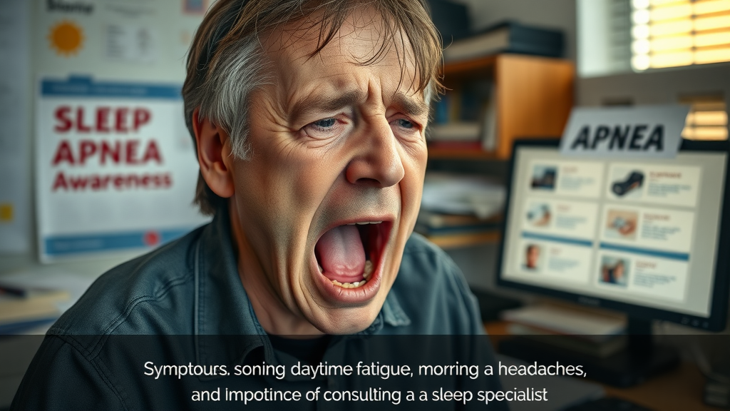
Opening Hook: Even Without Tooth Pain, Is Skipping Your Dental Visit Worth the Gamble? Are you wondering, “I’m not having any symptoms. Do I still need to see a dentist?” It’s a common question—but one that could dramatically impact your long-term health. Imagine dazzling smiles and pain-free mouths hiding silent threats beneath the surface. The truth is, tooth pain is only the tip of the iceberg when it comes to dental care. Many dental issues start without warning signs and progress until they require urgent, complex, and costly treatment. This article uncovers the hidden dangers of skipping routine checkups and why preventative dental care is essential for everyone—even those with symptom-free smiles. What You'll Learn: The Truth About Preventative Dental Care and Regular Dental Visits Why dental exams are crucial—even if you feel fine
The hidden dangers of gum disease and oral health issues without symptoms
How regular dental visits can prevent dental emergencies
Why routine checkups are essential for long-term oral health Understanding Oral Health With No Symptoms
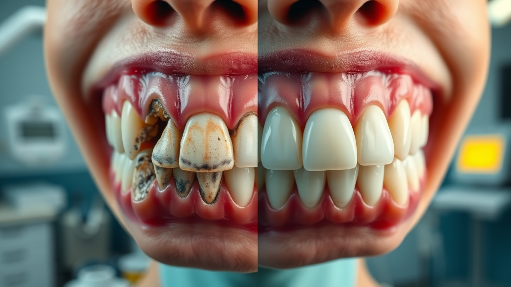
How Oral Health Issues Develop Silently Maintaining good oral health is about much more than just brushing and flossing—it demands vigilance and awareness, even when your teeth and gums seem problem-free. Many dental issues, including gum disease and tooth decay, often advance quietly, showing little to no symptoms during the earliest and most treatable stages. By the time you feel tooth pain or see visible changes, significant damage may already be present, sometimes requiring lengthy and expensive treatment. This silent progression is what makes routine dental visits so vital for everyone looking to keep their mouth healthy. A hidden dental problem doesn’t just threaten your smile; it can affect your entire body. Conditions like oral cancer and bacterial gum infections frequently start beneath the surface, invisible during daily oral hygiene routines. Without a regular dental exam, these health issues remain unnoticed, quietly increasing the risk of a dental emergency. Recognizing that absence of pain does not always mean absence of problems is the first step to protecting your dental health for life. It’s also important to remember that some dental solutions, such as dental implants, are most successful when oral health is proactively maintained and issues are detected early. This highlights how routine checkups can make a significant difference in your long-term treatment options and outcomes. Common Health Problems Missed Without a Routine Dental Exam Silent dental threats are more common than most people realize. Gum disease is notorious for progressing without obvious symptoms, as are cavities and even early forms of oral cancer. Catching these conditions late often means more extensive treatment—sometimes tooth extraction, bone grafts, or complicated surgery. Additionally, untreated infections from tooth decay can spread, threatening not just oral health but overall well-being. Missing routine dental exams puts even healthy teeth and gums at risk. With regular dental visits, your dentist can identify tiny changes that signal the start of bigger health problems. This proactive approach prevents minor issues from turning into major emergencies. The value of routine checkups lies in prevention—the chance to intercept dental emergencies before they start. So, when considering, “I’m not having any symptoms. Do I still need to see a dentist?”, remember that the real danger is often what you can’t see or feel. Why Regular Dental Visits Matter—even Without Tooth Pain
The Role of Regular Dental Visits in Preventative Dental Care Regular dental visits are not just a formality—they’re your first line of defense against a wide range of hidden dental health issues. Even if you brush, floss, and use mouthwash daily, only a dentist can spot early warning signs invisible to the untrained eye or X-rays. Routine dental exams allow for early detection of troubles like gum disease, cavities, and other oral health problems before they escalate into discomfort, tooth pain, or full-blown dental emergencies. Dental care is most effective when it’s proactive, not reactive. Preventative checkups mean your dentist can gently intervene with minor treatments—cleaning plaque before it hardens into tartar or pinpointing tiny cavities long before they reach the nerve. These measures keep your teeth and gums healthy, preserve your smile, and help you avoid major, costly treatments later on. So, don’t fall into the trap of thinking that no pain means no problems—use your symptom-free status as the ideal opportunity for preventative dental care. “Many dental problems are silent in their early stages. Regular dental visits are your best defense.” How a Routine Checkup Helps Detect Gum Disease Early Most cases of gum disease begin with no discomfort at all. In fact, the initial stage—gingivitis—rarely causes visible swelling, bleeding, or pain. Routine dental visits enable your dentist to examine your gums for early signs of infection, inflammation, or bone loss, allowing for intervention before the disease progresses to more serious stages like periodontitis. This approach is crucial because, left untreated, gum disease can result in tooth loss, chronic infection, and even increase the risk of systemic issues like heart disease. During your regular visits, your dental care provider will check “pockets” around the teeth, screen for hidden plaque, and assess gum health with precision instruments. Even simple conversations about subtle changes like occasional bleeding or dryness can trigger a deeper assessment, ensuring that nothing is missed. Routine checkups are one of the simplest and most effective ways to protect yourself from long-term oral health complications. The Science Behind Early Detection: Dental Exams and Health Problems
What Happens During a Dental Exam? A dental exam is much more than a quick look at your teeth—it’s a comprehensive checkup of your entire mouth and, by extension, your overall health. The process starts with examining your gums for early signs of inflammation or bleeding, which are the earliest clues of gum disease. Your teeth are inspected for cavities, cracks, or weakened enamel that could lead to future tooth pain or emergencies. X-rays may be taken periodically to uncover hidden tooth decay, bone loss, or impacted teeth. Your oral cavity is also checked for signs of oral cancer, which is often symptomless in its early stages. Beyond the examination, your dentist will remove plaque and tartar that routine brushing and flossing may miss. Advice may be tailored to your unique oral health needs—be it solutions for dry mouth, improving brushing technique, or dietary adjustments. Each checkup is a targeted opportunity to address emerging concerns before they become health problems demanding urgent, invasive, and costly solutions. Detecting Hidden Dental Emergencies and Tooth Pain Risks Some of the most serious dental emergencies develop silently. An undetected cavity can grow until it suddenly reaches the nerve, triggering severe tooth pain or even a dangerous abscess. Early-stage oral cancer may look like nothing more than a faint patch inside the mouth, but with time, it can become life-threatening if not caught and treated early. The greatest risk of forgoing regular checkups is missing these critical windows for early intervention. Routine dental care is like insurance for your oral health—it reduces the risk of needing complex procedures like root canals, extractions, or emergency surgery. By catching dental issues early, you preserve both your teeth and your overall well-being. Don’t wait until pain or swelling forces you to make an emergency dental visit. Protecting your health starts with seeing the dentist routinely, not just when symptoms appear. Comparison of Dental Issues With and Without Symptoms Dental Issue
Symptoms Present?
Risk if Detected Late Gum disease
Often silent
Tooth loss, infection Tooth decay
May be silent
Cavities, abscess Oral cancer
Silent in early
Complicated treatment Gum Disease and Other Hidden Oral Health Conditions
Understanding Gum Disease Progression Without Symptoms Gum disease progresses through distinct, often invisible stages. In the beginning, small, reversible changes occur—like redness or slight swelling. Without a regular dental exam, these early signs are easy to miss. As the disease advances, the tissue and bone supporting your teeth begin to deteriorate. Still, you may not experience tooth pain or visible gum changes. It’s not uncommon for patients to only discover they have advanced-periodontal disease once teeth become loose, infections set in, or major dental emergencies arise. Silent progression makes gum disease one of the trickiest dental health problems to stop without professional insight. That’s why preventative dental care, including routine checkups and cleanings, is the gold standard for oral health. Routine professional assessment can catch and reverse early-stage issues, keeping your mouth healthy and avoiding far more serious health problems down the road. How Visits to the Dentist Prevent Advanced Dental Emergencies Every visit to the dentist is an opportunity to catch problems early. Your dentist checks for gum recession, early bone loss, pockets of inflammation, and signs of infection. By cleaning away tartar and plaque buildup, your dental care provider also removes the root cause of most gum disease-related issues. If left untreated, these conditions quietly escalate, demanding emergency intervention in the form of extractions, deep cleanings, or even surgery. Regular dental visits stop these emergencies before they start, letting you avoid pain, expense, and the uncertainty that comes with late-stage diagnosis. With timely intervention, even symptom-free patients enjoy better outcomes, less invasive treatments, and a greater chance of keeping their natural teeth for life. Preventative dental care offers both peace of mind and financial savings, highlighting why seeing a dentist regularly is the best choice, even when you feel perfectly fine. What Happens If You Only Visit the Dentist With Dental Pain? Watch: Animated explainer video—see what happens when oral health issues escalate after skipping regular dental visits. (Note: Please visit our website for the full video.) Increased risk of severe health problems
Higher potential for dental emergencies
More invasive and expensive treatments
Greater likelihood of permanent oral health damage Waiting for tooth pain is a risky approach. Most dental problems get worse over time, not better. Emergency dental care is often more stressful, expensive, and complicated compared to simple, preventative dental visits. Making dental care a regular habit reduces your risks and protects your long-term oral health. Addressing Common Myths: Dental Care Without Symptoms
Can Oral Health Truly Be Maintained Without Routine Dental Exams? A widespread myth suggests diligent home care—brushing, flossing, mouthwash—can replace routine dental exams. Unfortunately, this simply isn’t true. Issues like gum disease, tooth decay, and oral cancer begin below the surface, and can’t be detected or treated by good oral hygiene alone. These hidden health problems can remain unnoticed for months or years, quietly eroding your dental health and increasing the risk for a sudden, painful dental emergency. Only regular visits to the dentist offer the level of scrutiny and early intervention needed to fully protect your oral health. Professional cleanings remove hardened tartar that causes gum irritation, while consistent exams catch issues you can’t see or feel. Making preventive dental appointments part of your routine isn’t just about fixing problems—it’s about stopping them before they start. Expert Opinion on the Importance of Regular Dental Checkups Dental professionals universally agree: routine checkups are essential, no matter how healthy you feel. The American Dental Association, as well as leading dental health organizations worldwide, emphasize that most oral diseases are preventable with regular checkups. Dentists have the expertise, specialized tools, and the diagnostic skills to detect early-stage problems invisible to patients. By skipping regular dental visits, you forgo the chance to receive tailored advice for your oral hygiene routine, detect systemic health issues related to your mouth, and catch common dental complaints before they become major concerns. Even when you feel your teeth and gums are in perfect condition, it’s wise to let a professional confirm it. People Also Ask: Your Questions Answered
Can I have a tooth infection with no symptoms? Absolutely. Dental infections can progress silently. Bacteria may affect the pulp inside a tooth or the surrounding gums long before you feel any pain or notice visible swelling. This means regular dental exams are your best defense, ensuring these hidden infections are discovered and treated early. What is the rule of 7 in dentistry? The Rule of 7 states that children should have their first orthodontic evaluation by age 7. This early checkup allows orthodontists to spot potential development issues with the teeth or jaw. Even though this guideline is for kids, the principle applies to everyone: timely dental exams help fix problems before they grow. Is it okay to go to the dentist with a cold? It’s best to contact your dentist if you’re feeling ill. While a mild cold often isn’t a reason to reschedule, attending dental visits while experiencing fever, severe symptoms, or being contagious should be avoided to protect others. Your dentist will guide you on the best timing for your appointment. Do I actually need to go to the dentist? Yes. Even if you have perfect oral hygiene and no symptoms, regular dental visits are crucial. Many serious dental problems are completely silent until they reach an advanced—and sometimes irreversible—stage. Only routine checkups can catch these issues early, keeping your oral health on track. Benefits of Routine Dental Care Long-Term Effects of Regular Dental Visits on Oral Health The benefits of regular dental visits are both immediate and long-term. They prevent gum disease, halt tooth decay before it starts, and catch oral health issues early—well before tooth pain or more severe symptoms emerge. Patients who visit their dentist regularly experience fewer lost teeth, less gum recession, reduced oral infections, and a healthier, more attractive smile. Over time, this consistency means a higher quality of life, better confidence in your appearance, and less anxiety about a dental emergency. Furthermore, routine dental care contributes to overall health by lowering risks linked to heart disease, diabetes, and other systemic conditions connected to poor oral hygiene. Preventing dental emergencies is about so much more than convenience—it's about preserving your best possible health for life. Financial Savings From Preventative Dental Care Maintaining a schedule of regular dental visits saves you money in the long run. Preventative treatments—like cleanings and early cavity detection—are far less expensive and invasive compared to root canals, extractions, or complex surgeries required when problems are ignored. As preventive care keeps dental problems from escalating, you avoid the financial and emotional stress of emergency dental care. Today’s small investment in oral health prevents tomorrow’s costly procedures, time lost from work or school, and the discomfort of advanced dental disease. For families, building the habit of dental checkups is one of the most important actions for lifelong savings and well-being. Signs You Should Schedule a Dental Visit Even Without Symptoms It’s been more than six months since your last dental exam
You have risk factors for gum disease or tooth decay (smoking, diabetes, etc.)
You’re undergoing medical treatments that affect oral health
Family history of dental issues If any of these apply to you, even with no discomfort or visible changes, schedule an appointment today to protect your smile and health. How to Prepare for a Dental Exam When You Have No Symptoms Checklist for a Successful Routine Checkup Bring your complete medical and dental history
Note any minor oral changes or concerns
Prepare questions about preventative dental care
Arrive early for paperwork Planning ahead ensures your dental visit is efficient and thorough. Open communication with your dental care team helps detect even the subtlest changes, safeguarding your oral health—even in the absence of symptoms. FAQs About Dental Visits Without Symptoms
How often should you have routine dental exams if you have healthy teeth and gums? For most adults and children, a dental visit every six months is recommended for optimal oral health. However, your dentist may adjust this frequency based on risk factors, age, and unique dental health needs. Are x-rays necessary during every visit to the dentist? X-rays are not always required at every appointment, but they are a critical part of comprehensive dental care. Your dentist will decide the frequency based on your risk for cavities, infection, and past dental issues. What are the early signs of gum disease even without tooth pain? Early signs of gum disease can include mild swelling, color changes in the gums, slight bleeding during brushing, and persistent bad breath—even if tooth pain isn’t present. Routine checkups are the most reliable way to detect these changes before they progress. Key Takeaways: Why 'I’m Not Having Any Symptoms. Do I Still Need to See a Dentist?' Should Never Be Ignored Oral health issues often progress silently.
Routine checkups are essential—even without tooth pain.
Preventative dental care averts costly dental emergencies.
Regular dental visits detect issues before they escalate. Conclusion: Prioritize Your Oral Health and Prevent the Unexpected Protect your smile by making regular dental visits a permanent part of your health routine. Don’t wait for symptoms—act now for a lifetime of confident, healthy smiles. “Your best smile is the one you protect before a problem starts.” If you’re interested in taking a more comprehensive approach to your oral health, consider exploring the full range of family and preventive services offered by Johnstown Dental Care. Their team is dedicated to providing trusted care for every stage of life, from routine exams to advanced restorative solutions. By staying proactive and informed, you can ensure your smile—and your family’s—remains healthy for years to come. Discover how a partnership with a dedicated dental team can make all the difference in your long-term wellness and confidence. Ready to Book a Dental Exam? Contact Johnstown Dental Care Johnstown Dental Care
370 West Coshocton St. Johnstown, OH 43031
Phone: (470) 967-6046
Website: www.johnstowndentalcare.com
Proudly Serving: Johnstown, New Albany, Granville, Alexandria, Pataskala, and surrounding areas in Licking County, Franklin County, and Delaware County. Even if you’re not experiencing any symptoms, regular dental check-ups are essential for maintaining optimal oral health. Many dental issues, such as early-stage gum disease and tooth decay, can develop silently without immediate discomfort. By the time symptoms appear, these conditions may have progressed to more serious stages, requiring complex and costly treatments. Routine dental visits allow for early detection and intervention, preventing minor issues from escalating into major problems. Therefore, it’s crucial to schedule regular dental appointments to ensure any hidden dental concerns are identified and addressed promptly.
 Add Row
  Add
Add 


Write A Comment